
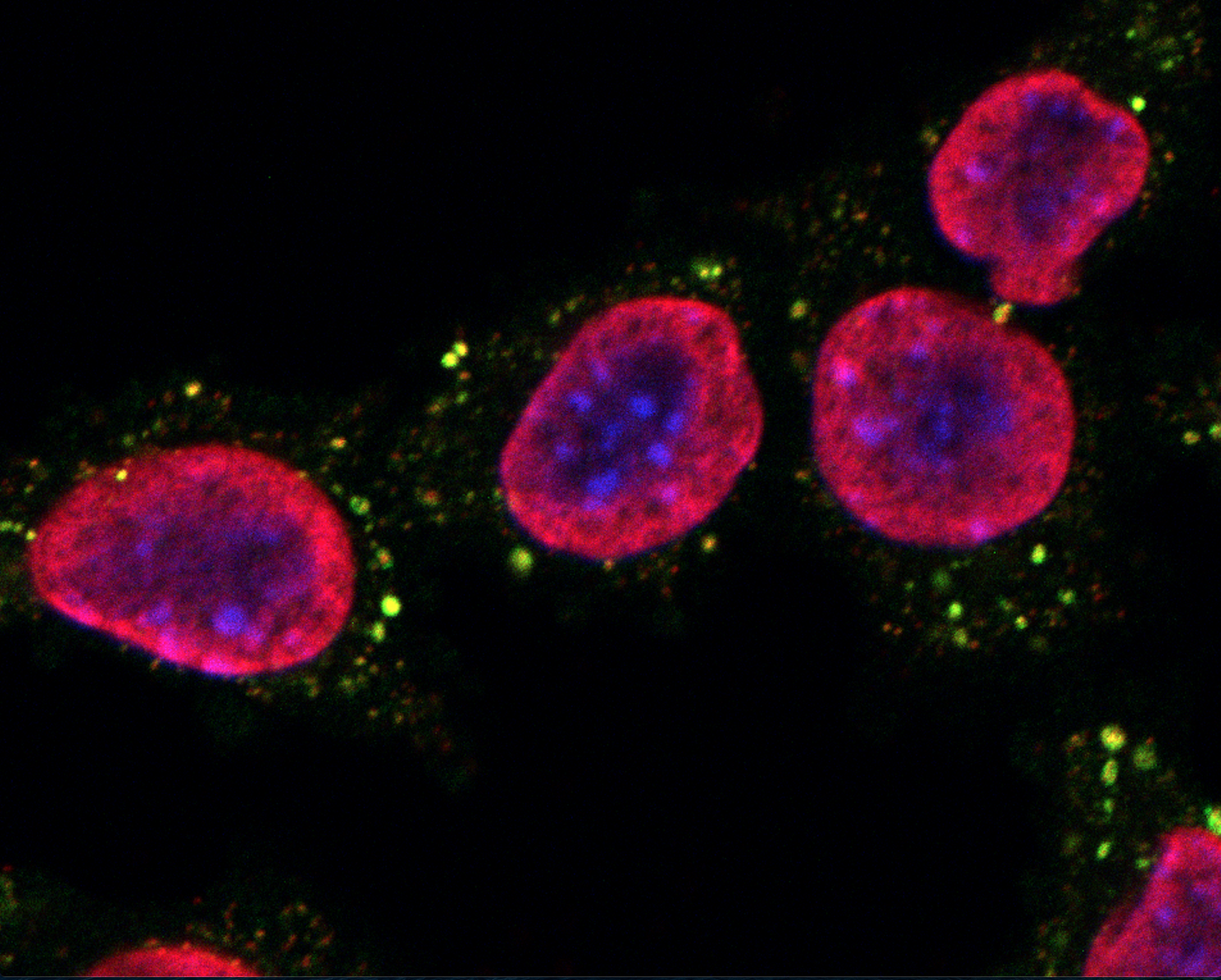
Mouse breast carcinoma TSA cells maintained in control conditions and then subjected to strong permeabilization followed by staining with antibodies specific for dsDNA (red) and TFAM (green). DAPI (blue) was used for nuclear counterstaining. High magnification. Courtesy of Ai Sato (© Ai Sato, Lorenzo Galluzzi).
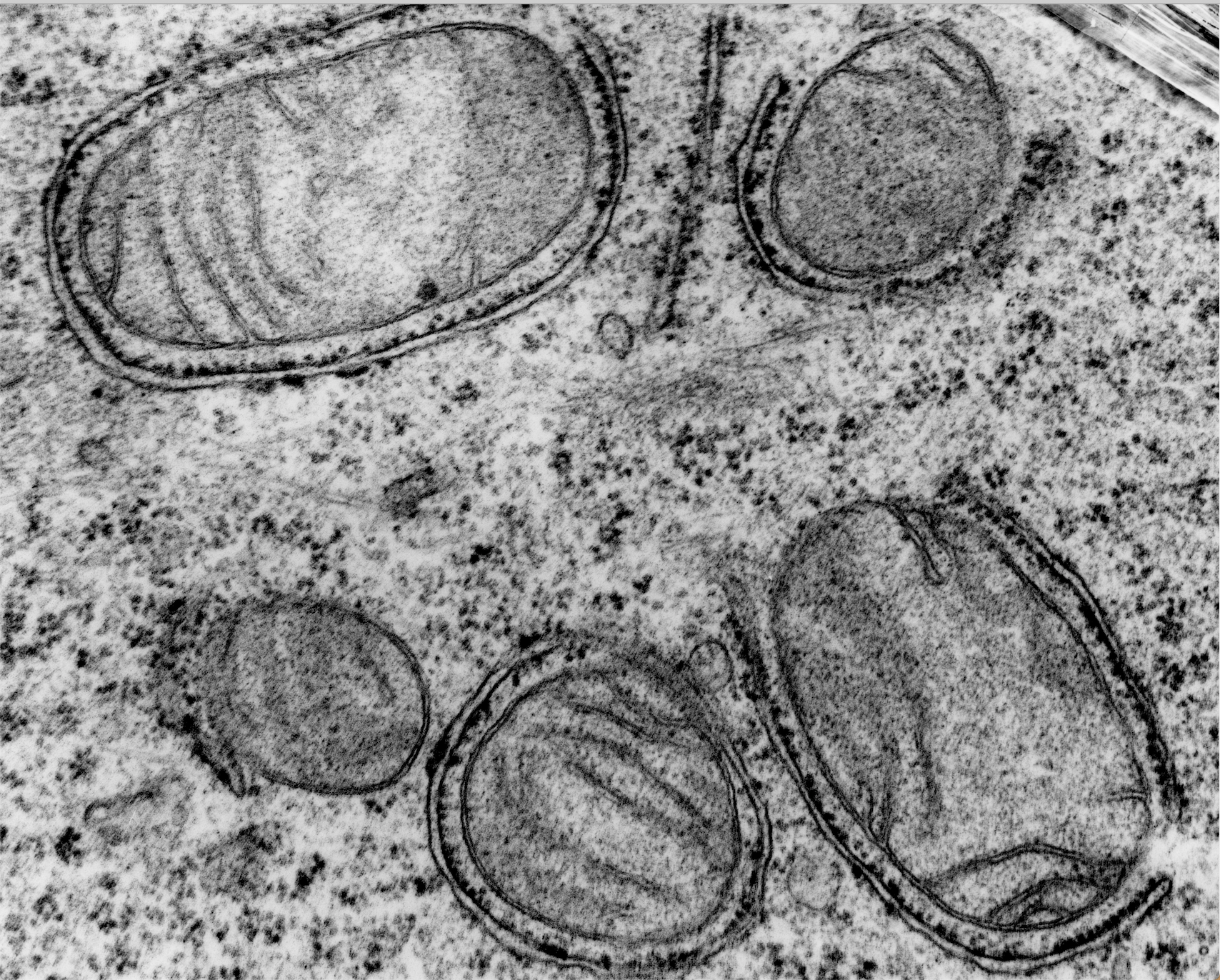
Autophagosomes surrounding mitochondria with partial cristae disruption in a human non-small cell lung carcinoma H1975 cell. Courtesy of Gerard Pierron (©Gerard Pierron, Lorenzo Galluzzi).
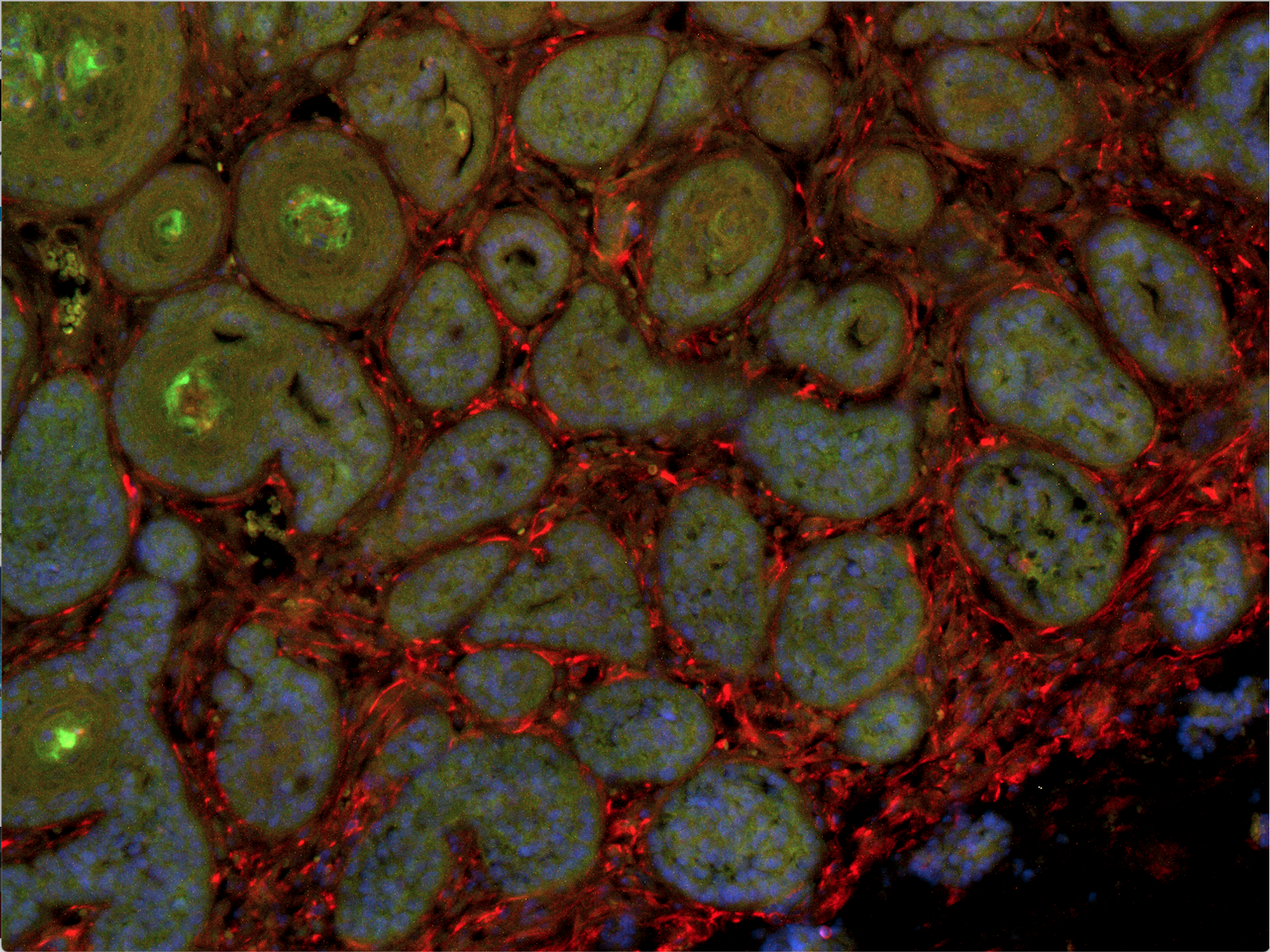
MPA/DMBA-driven tumor evolving untreated in a C57BL/6 mouse and stained for estrogen receptor 1 (ESR1, green) and vimentin (VIM, red) expression. DAPI (blue) was used for nuclear counterstaining. Low magnification. Courtesy of Ai Sato (© Ai Sato, Lorenzo Galluzzi).
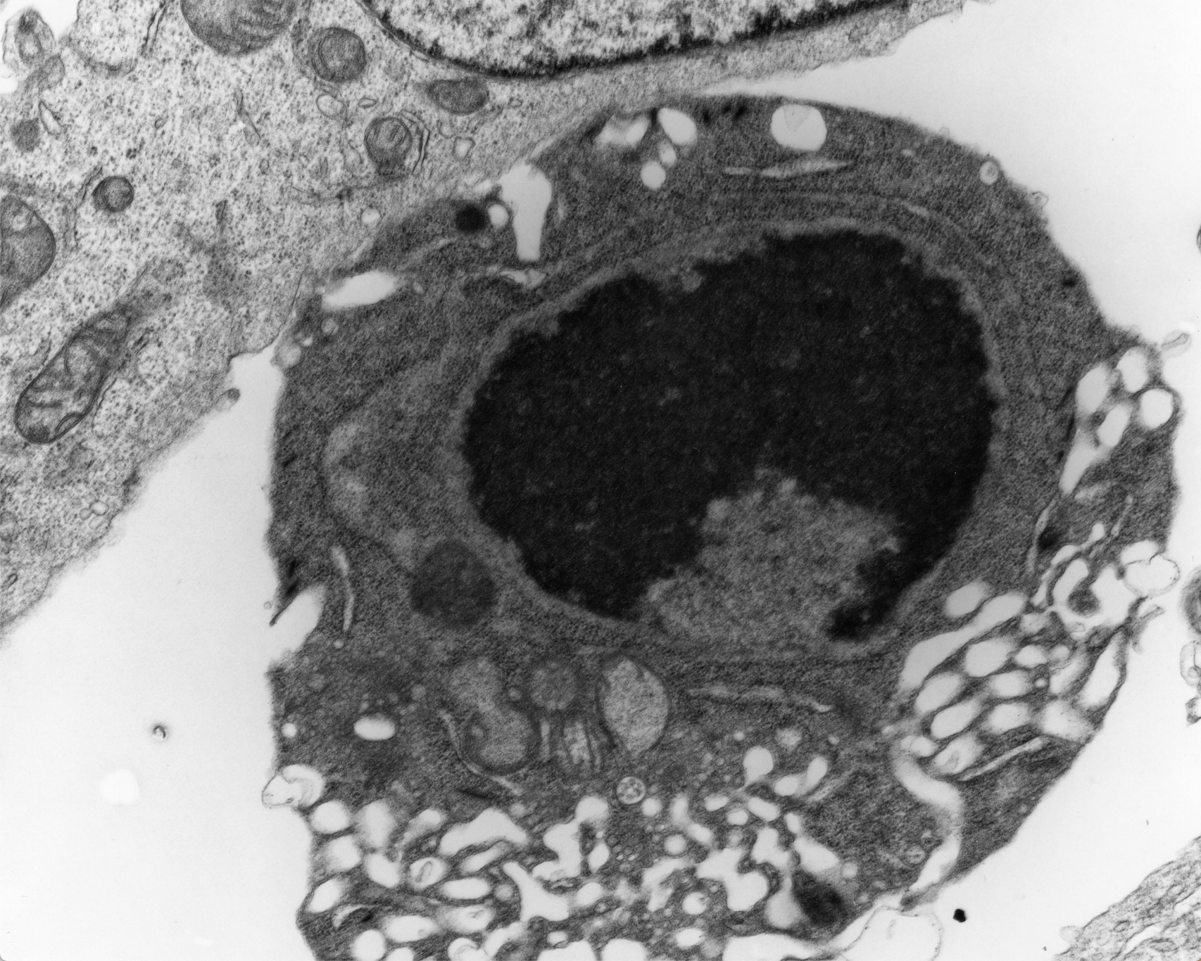
Apoptotic human non-small cell lung carcinoma H1975 cell. Courtesy of Gerard Pierron (©Gerard Pierron, Lorenzo Galluzzi).
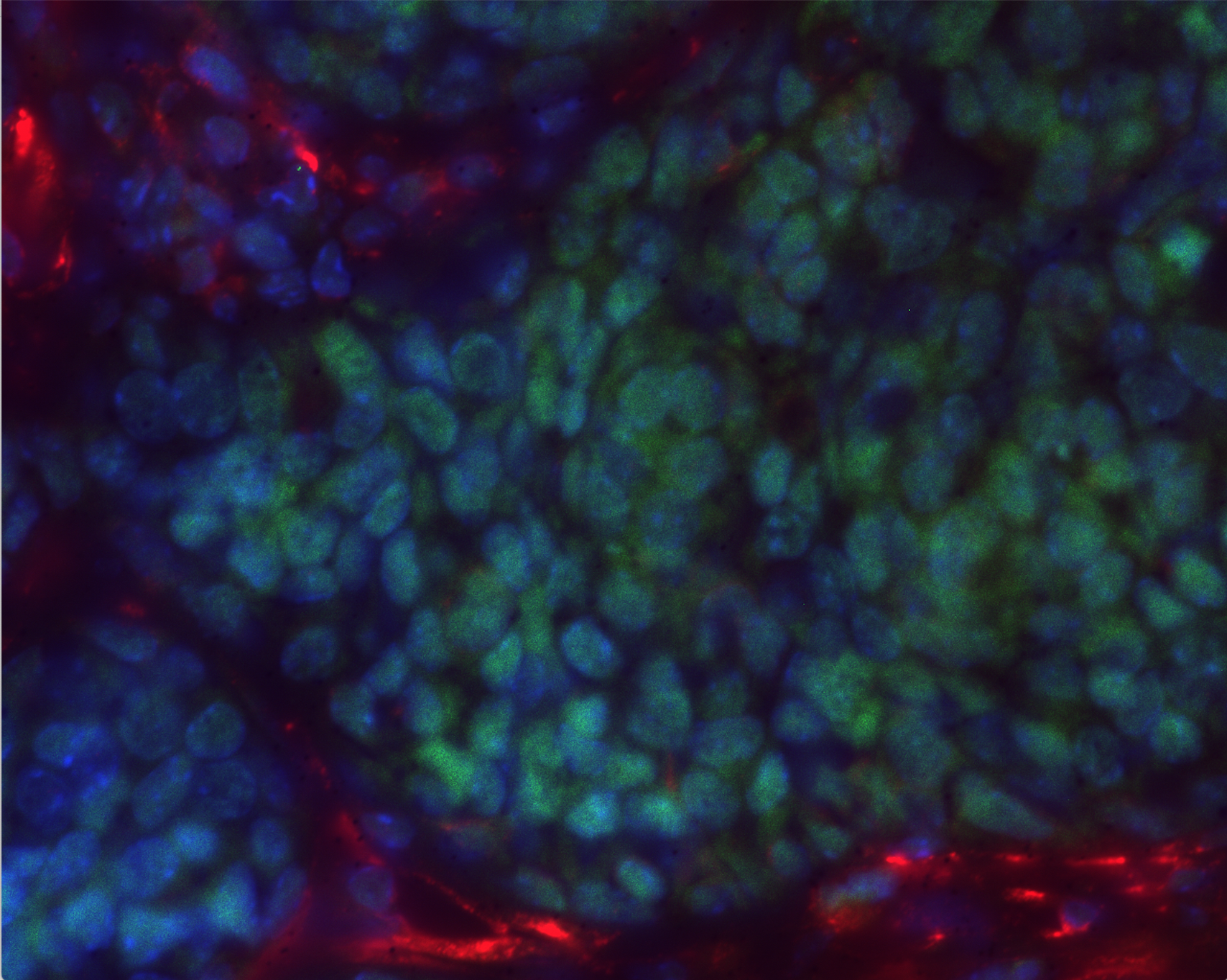
MPA/DMBA-driven tumor evolving untreated in a C57BL/6 mouse and stained for estrogen receptor 1 (ESR1, green) and vimentin (VIM, red) expression. DAPI (blue) was used for nuclear counterstaining. High magnification. Courtesy of Ai Sato (© Ai Sato, Lorenzo Galluzzi).
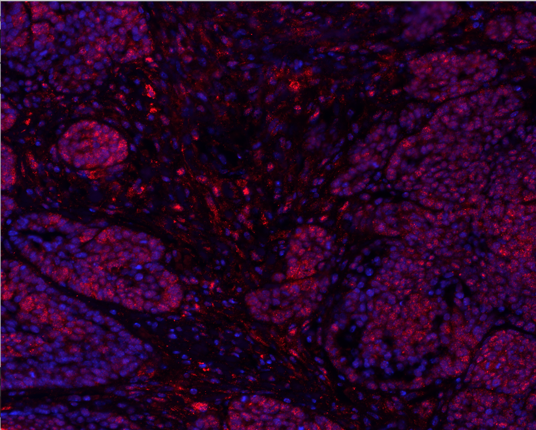
MPA/DMBA-driven tumor evolving untreated in a C57BL/6 mouse and stained for three prime repair exonuclease 1 (TREX1, red). DAPI (blue) was used for nuclear counterstaining. High magnification. Courtesy of Ai Sato (© Ai Sato, Lorenzo Galluzzi).
Give your help to cancer research.
Click here to make a secure gift by credit card.
With your support, we can make the difference.